National Institutes of Health scientists have used human skin cells to create what they believe is the first cerebral organoid system, or “mini-brain,” for studying sporadic Creutzfeldt-Jakob disease (CJD). CJD is a fatal neurodegenerative brain disease of humans believed to be caused by infectious prion protein. It affects about one in one million people. The researchers, from NIH’s National Institute of Allergy and Infectious Diseases (NIAID), hope the human organoid model will enable them to evaluate potential therapeutics for CJD and provide greater detail about human prion disease subtypes than the rodent and nonhuman primate models currently in use.
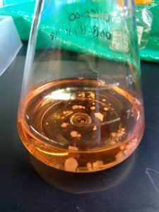
Human cerebral organoids are small balls of human brain cells ranging in size from a poppy seed to a small pea. Their organization, structure, and electrical signaling are similar to brain tissue. Because these cerebral organoids can survive in a controlled environment for months, nervous system diseases can be studied over time. Cerebral organoids have been used as models to study Zika virus infection, Alzheimer’s disease, and Down syndrome.
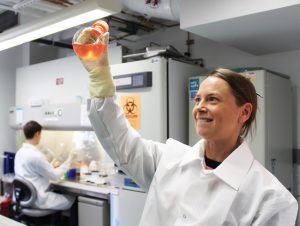
In a new study published in Acta Neuropathologica Communications, scientists at NIAID’s Rocky Mountain Laboratories in Hamilton discovered how to infect five-month-old cerebral organoids with prions using samples from two patients who died of two different CJD subtypes, MV1 and MV2. In an interview at the laboratory, Dr. Cathryn Haigh, team leader on the mini-brain project, was excited about the recent results.
Dr. Haigh came to RML two years ago from Lebourne, Australia, where she had begun developing the idea of possibly using cultured brain tissue to study prion diseases.
“We didn’t invent this technique of growing cultured brain tissue,” Said Dr. Haigh. “Our contribution was in creating this prion infection model by demonstrating that a prion disease can be transmitted to human brain cells.”
One part of the process that Dr. Haigh appreciates is that they don’t need to start with any human brain cells. They make them. All the participating patient has to contribute is a small sample of skin. These skin cells are then “genetically engineered,” reducing them to stem cells and then stimulating the development of brain cells out of that stem component by administering certain hormones.
The result is a bunch of tiny globs of brain tissue, little mini-brains, floating in a glass flask. The scientists began by adding CJD infected tissue to the fluid in which the mini-brains float, exposing the organoids to the disease and then removing it. Then they observed and monitored for signs of infection. They used two basic bio-chemical methods of prion detection and within about a month confirmed that the mini-brains had been infected. The mini-brains were further monitored to track the course of the infection by checking certain health indicators such as levels of metabolism and electrical activity. By the end of the study, the scientists observed that seeding activity, an indication of infectious prion propagation, was present in all organoids exposed to the CJD samples. However, seeding was greater in organoids infected with the MV2 sample than the MV1 sample. They also reported that the MV1-infected organoids showed more damage than the MV2-infected organoids.
Dr. Haigh called discerning the difference between the two sub-types “intriguing.”
“We know that in human patients they behave differently,” she said. “One kills faster than the other one and the patients display different symptoms.” She said this indicates that they are attacking different cell populations in the brain. “Now, hopefully, we can see what types are being attacked and find out why and possibly stop it.”
She said they plan to further investigate those differences in hopes of identifying how different subtypes of CJD affect brain cells. Ultimately, they hope to learn how to prevent cell damage and to restore the function of cells damaged by prion infection. The new system also provides opportunities to test potential therapeutics for CJD in a tissue model that mimics the human brain.

To date, most pharmaceutical research for drug treatments has been based on experiments on mice and non-human primates. One big issue in the industry, she said, has been that many of the successful studies on mice don’t show the same results when finally tested on humans. By using cerebral organoids, scientists can get one step closer to determining the potential results on humans without actually testing it on a human.
Dr. Haigh is aware that there are limitations to the model. The organoids lack a vascular system and some of the other cells common throughout the brain.
“Despite these limitations,” she said, “these cultures represent the closest in vitro cell model to human brain currently available and offer the first human three-dimensional model of prion disease.” Their use holds promise, she said, for the investigation of different subtype pathologies, for investigation of how prions spread throughout the brain and for trialing putative therapeutics in a human tissue background.